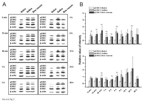
Figure 2.

Temporal changes in phosphorylation and expression of ERKs in contralateral S1 area under three different states. (A) Representative Western blots of neocortical homogenates from three groups of rats at indicated time points. Left panel in each rectangle represents immunodetection of phosphorylated-ERK1/2 (pERK1/2), total ERK1/2 (tERK1/2), and beta-actin, which is included as loading control. Right panel in each rectangle shows raw bands corresponding to the left panel. (B) Densitometric quantification from (A) is shown. The pERKs bands were densitized and normalized to tERKs immunoblotted from the same membrane to demonstrate the phosphorylation levels of ERKs. Upper panel of B denotes the time course of ERK1 activation under three assigned states, while lower panel of B illustrates the time-related changes in activation of ERK2. Points represent the mean ± S.E.M. from three separate experiments. * P < 0.05, ** P < 0.01, saline-treated rats versus naïve control. † P < 0.05, †† P < 0.01, bee venom-treated rats versus naïve control.
