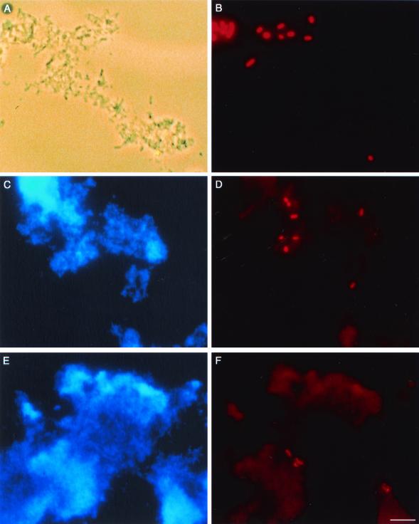
FIG. 3.
Micrographs of E. coli in 2-week-old biofilms. Total bacteria were visualized using phase-contrast microscopy (A) or CPI staining (C and E). E. coli was detected in the same fields after hybridization with the rhodamine-labeled 16S rRNA-targeted oligonucleotide Eco3 probe (B, D, and F). E. coli was inoculated into biofilms 8 and 10 and detected 10 days later, after exposure to 1 CT hypochlorous acid (A and B) or 4 ppm NH2Cl for 155 min (C and D). E. coli present in the Albany distribution system was detected in biofilm 4, exposed to 1 CT hypochlorous acid (E and F). Bar, 5 μm.

