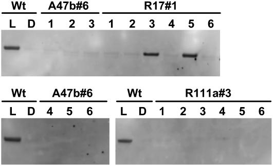
Figure 2.
Immunodetection of phosphorylated PEPC in leaf extracts of F. bidentis wild type (Wt) and progeny of T1 transformants A47b#6, R111a#3, and R17#1 shown in Figure 1. Proteins were separated by SDS-PAGE. Plants were grown in a naturally lit glasshouse during June and July, 2006, and leaf discs were collected in the glasshouse for wild type either during the dark period (D) or at midday (L). Leaf samples for the progeny of T1 transformants were collected at midday in the light. (Samples were loaded on gels on an equal-leaf-area basis.) Phosphorylated PEPC was detected with the specific antibody and with anti-IgG alkaline phosphatase conjugate (Bio-Rad) as secondary antibody. Immunoblots were developed using the AttoPhos fluorescence substrate system (Promega). There were no other bands on the membrane.

