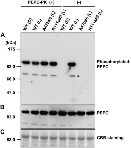
Figure 4.
In vitro rephosphorylation of PEPC. The left-hand side shows the phosphorylation state of PEPC extracted form leaves of wild type (Wt) collected at midnight in the dark (D) or collected at midday in the glasshouse in the light (L) and from progeny of A47b#6 and R111a#3 collected at midday in the light after in vitro phosphorylation of PEPC in the presence of recombinant PEPC-PK from F. trinervia. A, Phosphorylated PEPC was detected with the specific antibody and with anti-IgG HRP conjugate (Pierce) as secondary antibody. Immunoblots were developed using the chemiluminescent HRP substrate system (Millipore) and visualized with the luminoimage analyzer LAS-1000 (Fuji Film). The right-hand side shows the phosphorylation state of PEPC in the same leaf extracts without the addition of recombinant PEPC-PK (for details, see “Materials and Methods”). B, PEPC levels were also immunologically detected by reprobing the same membrane using anti-maize C4 PEPC antibody (for details, see “Materials and Methods”). C, A duplicate gel was run and stained with Coomassie Blue. On the left side on each image, positions of protein size makers are indicated. An asterisk indicates additional signals, solely observed in the rephosphorylation experiment due to longer incubation times.

