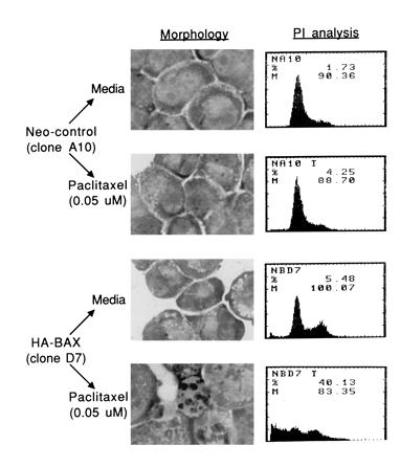
Figure 4.

Effects of HA-BAX on morphology and sub-G0 content of SW626 cells treated with paclitaxel. Neo-control clone A10 and HA-BAX clone D7 were grown for 48 hr in the presence of medium alone or paclitaxel (0.05 μM), followed by morphologic assessment (Wrights–Giemsa staining) and quantitation of the sub-G0 fraction by propidium iodide staining as described. Significant amounts of nuclear fragmentation and an increased fraction of sub-G0 cells are observed for HA-BAX clone D7 compared with neo-control cells, consistent with paclitaxel-mediated apoptosis. Representative data from one of three separate experiments are shown.
