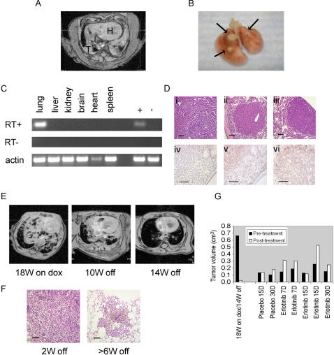
Figure 6. Characteristics of C/T790M mice.
A. Axial MR image from a line 37-derived bitransgenic animal fed dox for 32 weeks, revealing tumor (T); H: heart. B. Gross histology shows corresponding lung lesions. C. RT-PCR was performed on lungs from this animal in the presence or absence of RT using transgene-specific primers on mRNA from various tissues; “+”–positive control; “−“– negative control. D. Images of H&E-stained lung sections from various T790M transgene bearing mice. Lungs from bitransgenic line 37 (i) and 8 animals (ii-iii) displayed features of papillary adenocarcinoma (i), solid adenocarcinoma surrounded by bronchioloalveolar carcinoma (ii), and solid/papillary adenocarcinoma surrounded by bronchioloalveolar carcinoma (iii). Tumors were negative for CC26 (iv), positive for SP-C (v), and positive for phospho-EGFR (Y1016) (vi). Bars, 100 microns. E. Serial MR images of a bitransgenic C/T790M animal (line 8) administered dox 18 weeks (left) and then fed a normal diet for the indicated times; W–weeks. F. H&E-stained sections from lungs of bitransgenic tumor-bearing mice withdrawn from dox for the indicated times. Left–degenerating tumor with scattered viable cells; Right–hemosiderin-laden macrophages engulfing a degenerating tumor. G. Tumor volumes were quantitated in the MR images obtained from the individual mice pre- and post-treatment with placebo or erlotinib. See Table S2 and methods for details. W–weeks, D–days.

