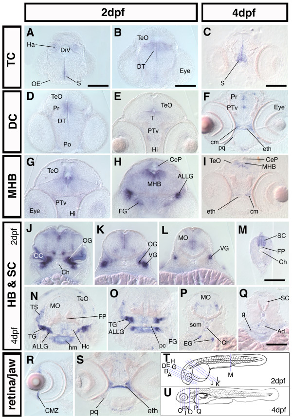
Figure 4.
nestin is expressed in proliferative zones during the development of the CNS. Cross sections of 2 dpf and 4 dpf embryos; dorsal up. (A-S): frontal and transversal cross section of nestin expression along the rostrocaudal axis of the brain and in the spinal cord reveals expression in major proliferative zones The relative rostrocaudal levels of each section are indicated in T and U. (A-I): sections at levels of forebrain and midbrain as well as midbrain-hindbrain boundary. (J-Q): sections at the level of HB and SC, showing nestin expression in the cranial ganglia and the HB, MO, SC. (R): cross section through the eye of a 4 dpf embryo, nestin is expressed in the CMZ; (S): cross section of a 4 dpf embryo at the level of the ethmoid plate, nestin is expressed in the craniofacial mesenchyme adjacent to developing cartilage tissue. (T, U): scheme of a 2 dpf (T) and a 4 dpf (U) zebrafish embryo, the levels of the cross sections shown in A-S are indicated by blue lines. Abbreviations: Ad: Aorta dorsalis; ALLG: anterior lateral line ganglion; CeP: cerebellar plate; Ch: chorda dorsalis; cm: craniofacial mesenchyme; CMZ: ciliary marginal zone; DiV: diencephalic ventricle; DT: dorsal thalamus; eg: enteric ganglia; eth: ethmoid plate; FG: facial ganglion; FP: Floor plate; g: gut; Ha: habenula; Hc: caudal hypothalamus; Hi: intermediate hypothalamus; hm: head mesenchyme; MHB: midbrain hindbrain boundary; MO: medulla oblongata; OC: otic capsule; OE: olfactory epithelium; OG: octaval ganglion; pc: parachordal cartilage; pq: palatoquadrate; Po: preoptic region; Pr: pretectum; PTv: ventral part of the posterior tuberculum; S: subpallium; SC: spinal cord; som: somites; T: midbrain tegmentum; TeO: optic tectum; TG: trigeminal ganglion; TS: torus semicircularis; VG: vagal ganglion. Scale bars: 100 μm.

