
Figure 1. Expression of ppk-1 in the adult hermaphrodite.
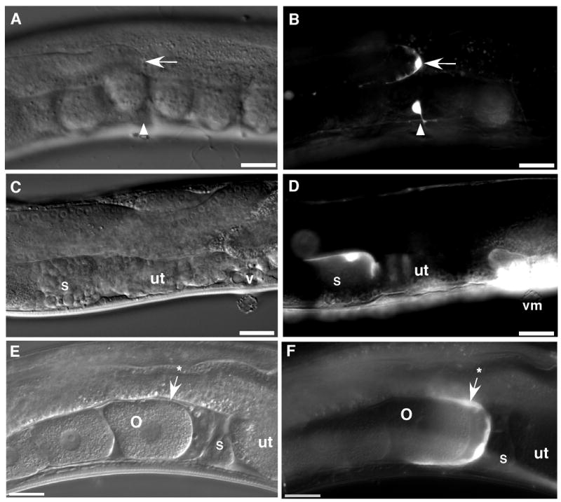
ppk-1::GFP transgenic animals were visualized by DIC and fluorescence microscopy. Bright field DIC images (panels A, C, and E) show the mid-body of a representative transgenic C. elegans hermaphrodite, including, panel A: distal tip (arrow) of the gonad and ventral mid-body (arrowhead), panel C: spermatheca (s), uterus (ut), and vulva (v), and panel E: oocyte (o), spermatheca (s), and gonad sheath (arrow with an asterisk). Fluorescence images (panels B, D, and E) of the same animals indicate that ppk-1::GFP is expressed in the distal tip cell (arrow), ventral neuron (arrowhead), spermatheca (s), vulval muscles (vm), and gonad sheath (arrow with an asterisk). Bars = 20 μm.