
Figure 4. Proximal gonads of ppk-1 (RNAi) animals.
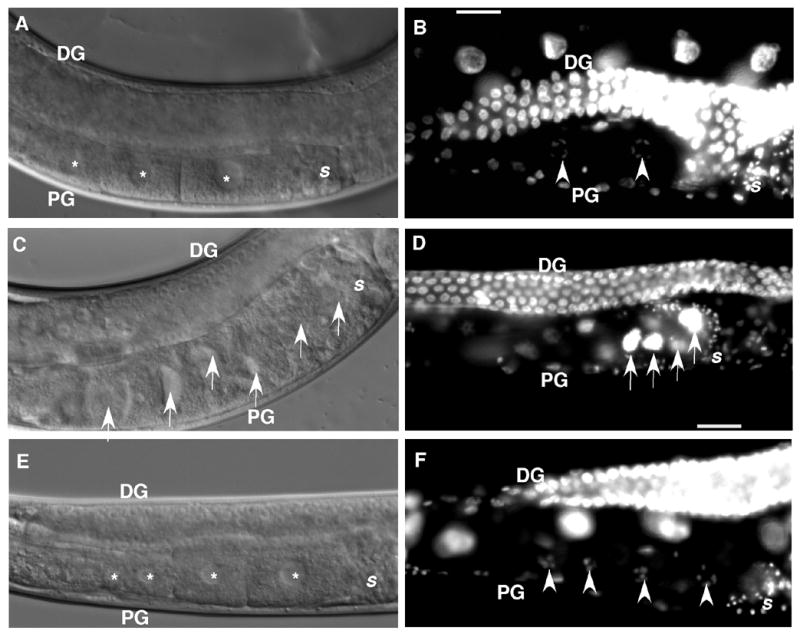
Panels A, C, and E: DIC microscopy images. Panels B, D, and F: DAPI stained images. Panels A and B: N2; control RNAi worm. A normal assembly line of developing oocytes (panel A, asterisk) in the proximal arm of the gonad (PG) and typical diakinetic oocyte nuclei (panel B, arrowheads) are visualized. Panels C and D: Control N2 worms treated with ppk-1 (RNAi). Oocytes accumulate (arrows) in a non-linear arrangement. The proximal gonad of N2; ppk-1 (RNAi) animals is filled with endomitotic (Emo) oocyte nuclei (panel D, arrows). Panels E and F: ppk-1 (RNAi) in the itr-1 (sy290) background. The gain-of-function itr-1 rescues oocyte accumulation. Developing oocytes display a typical linear arrangement (panel E, asterisk) in the proximal gonad. DAPI staining reveals no evidence of Emo oocytes and shows an organized linear arrangement of diakinetic nuclei (panel F, asterisk). DG: distal gonad. PG: proximal gonad. S: spermatheca.