
Figure 1.
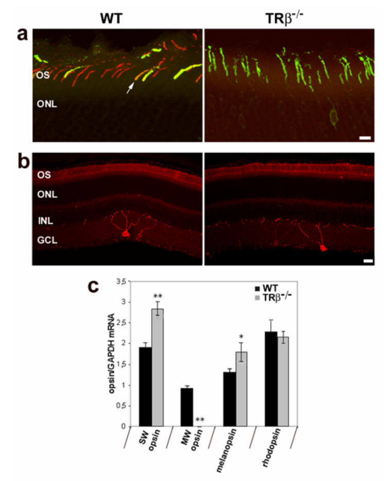
Cone opsins and melanopsin expression in the retina of wild-type (WT) and MW-coneless (TRβ−/−) mice, a) Confocal photomicrographs showing immunohistochemical labelling of MW (red fluorescence) and SW (green fluorescence) opsins in retinal sections. In wild-type mice, both MW and SW opsins are present with co-expression of both opsins in the outer segments of some cones (white arrow, yellow fluorescence). In TRβ−/− mice, MW-immunoreactive cones are not detected and all cones express SW opsin. Scale = 10μm b) Confocal photomicrographs of melanopsin-immunopositive ganglion cells (red fluorescence) in retinal sections of WT and MW-coneless mice showing that the relative number and distribution of melanopsin-containing ganglion cells are similar for both genotypes. Scale = 20 μm; OS: outer segment; ONL: outer nuclear layer; INL: inner nuclear layer; GCL: ganglion cell layer, c) Relative Opsins (SW, MW and rhodopsin) and melanopsin mRNA levels in the retina of WT (black bars) and MW-coneless (grey bars) mice using real time PCR. Results are expressed as mean ± SEM (n = 4 for each genotype). The TRβ−/− knockout mouse is characterized by a total absence of MW opsin and over-expression of SW opsin. The relative level of melanopsin is also up-regulated whereas rhodopsin levels are equivalent in both genotypes. Asterisks indicate a statistically significant difference (Mann-Whitney U test *:p<0.05; **: p<0.01).