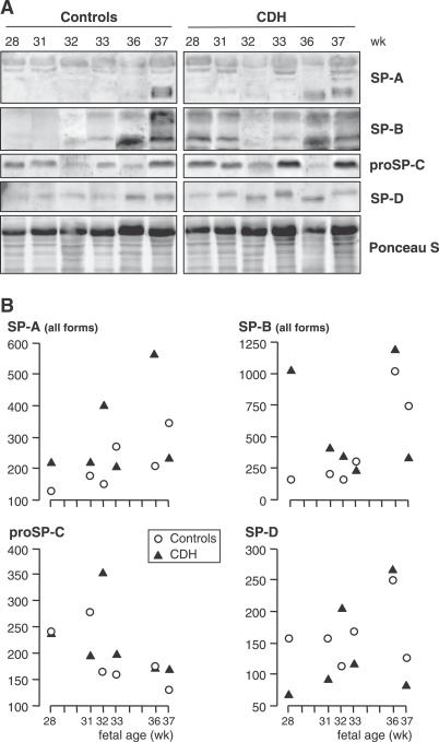
Figure 2. SPs in Human Fetal Lungs.
(A) Western blots: Samples were electrophoresed by SDS-PAGE, transferred, and successively incubated with specific anti-SP antibodies. (B) Densitometric analysis normalized by Ponceau S for gel loading (arbitrary units). SPs were detected at all stages. SP-A monomers were extremely faint before 36 wk, when they increased sharply (A) resulting in an increase in total amount (B). SP-B also increased markedly at 36–37 wk, ProSP-C did not display developmental changes, and SP-D showed a weak increase (A, B). There were no obvious differences between CDH and control lungs for any SP, with developmental changes occurring at the same fetal ages, the only exception being presentation of a high SP-B level in one CDH fetus, aged 28 wk.

