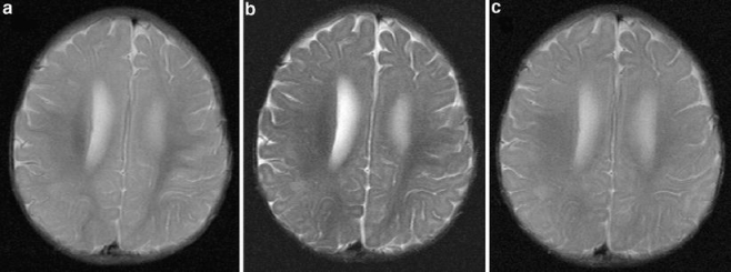
Fig. 1.

Axial images acquired through the bodies of the lateral ventricles of a 6-month-old child. First (a) and second (b) echo (TE 30/120 ms, TR 5,500 ms, TI 130 ms) of a dual-echo short-tau sequence shows the increased contrast between grey and white matter on the second echo compared to (c) the T2-W (TE 90 ms, TR 3,500 ms) sequence that would be used in those over 2 years of age. The slice thickness (5 mm) and matrix size (512 × 192) were the same for both sequences
