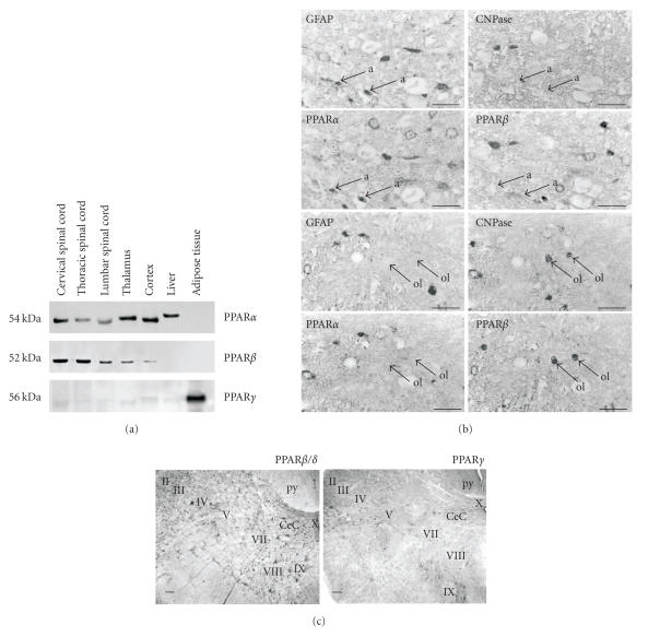
Figure 2.
Localization of PPARα and PPARβ/δ in astrocytes and oligodendrocytes in the spinal cord. (a) Western blotting shows PPARα and PPARβ but not PPARγ in spinal cord, telencephalon, and diencephalon. Identical expression patterns were detected with RT-PCR. (b) Detection of PPAR immunoreactive cells in the white matter of rat spinal cord. GFAP-positive/CNPase-negative astrocytes are immunoreactive for PPARα (marked a, upper four panels) while GFAP-negative/CNPase-positive oligodendrocytes express PPARβ (marked ol, lower panels) (scale bars: 25μ m, source: [38]). (c) Distribution of PPARβ/δ and PPARγ immunoreactive cells in the spinal cord of the adult rat (cervical level, coronal sections, scale bar: 100μ m, source: [41]).

