
Figure 6.
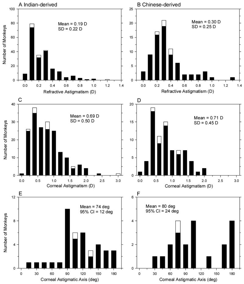
Distributions of refractive astigmatism (A & B), corneal astigmatism (C & D), and the corneal astigmatic axes (E & F) for the Indian- (left) and Chinese-derived monkeys (right) at 3 weeks of age. The cross-sectional and longitudinal groups are represented by the filled and open bars, respectively.