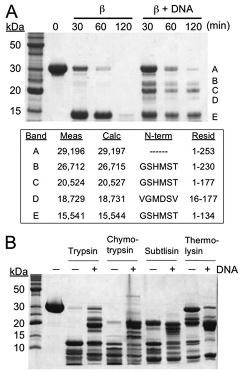
FIGURE 1. Limited protease digestion of β protein in the presence and absence of DNA.

The digestion was performed on β protein alone or in complex with DNA as formed by the sequential addition of two complementary 33-mer oligonucleotides. A, time course of limited trypsin digestion. The text box shows for each of the major fragments the mass measured by LC-MS, the mass calculated from the amino acid sequence, the results from N-terminal sequencing, and the corresponding residues of β protein. Notice that formation of the DNA complex confers protease resistance to residues 135–230 of β protein, as seen by the appearance of bands B, C, and D. B, limited digestion with trypsin, chymotrypsin, subtilisin, and thermolysin for 30 min in the presence and absence of DNA. The identities of each of the fragments produced were determined by mass spectrometry and are presented in supplemental Fig. S2.
