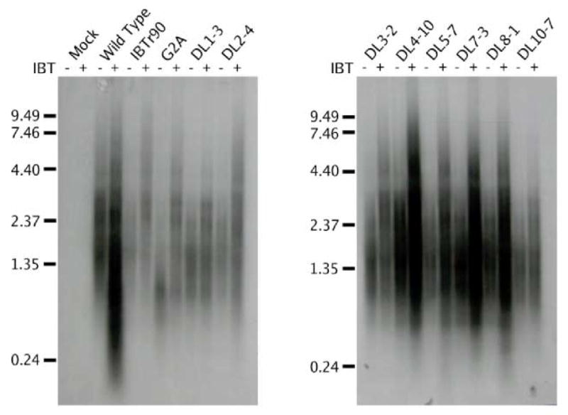
Fig. 5. Northern analysis of IBT-resistant mutant RNA.

Confluent BSC40 cells were infected with the indicated virus at m.o.i. = 15 in the presence or absence of IBT and incubated at 37°C for 9 hours. Total cellular RNA was purified, fractionated by gel electrophoresis and transferred to a nylon membrane. The membrane was probed with a riboprobe specific for the intermediate K2L gene and exposed to film. The virus used for infection is indicated at the top of the autoradiogram. The presence of absence of IBT in the infection is indicated with a “+” or a “−” at the top of the autoradiogram. Size markers are indicated at the left of the autoradiagram, in kb.
