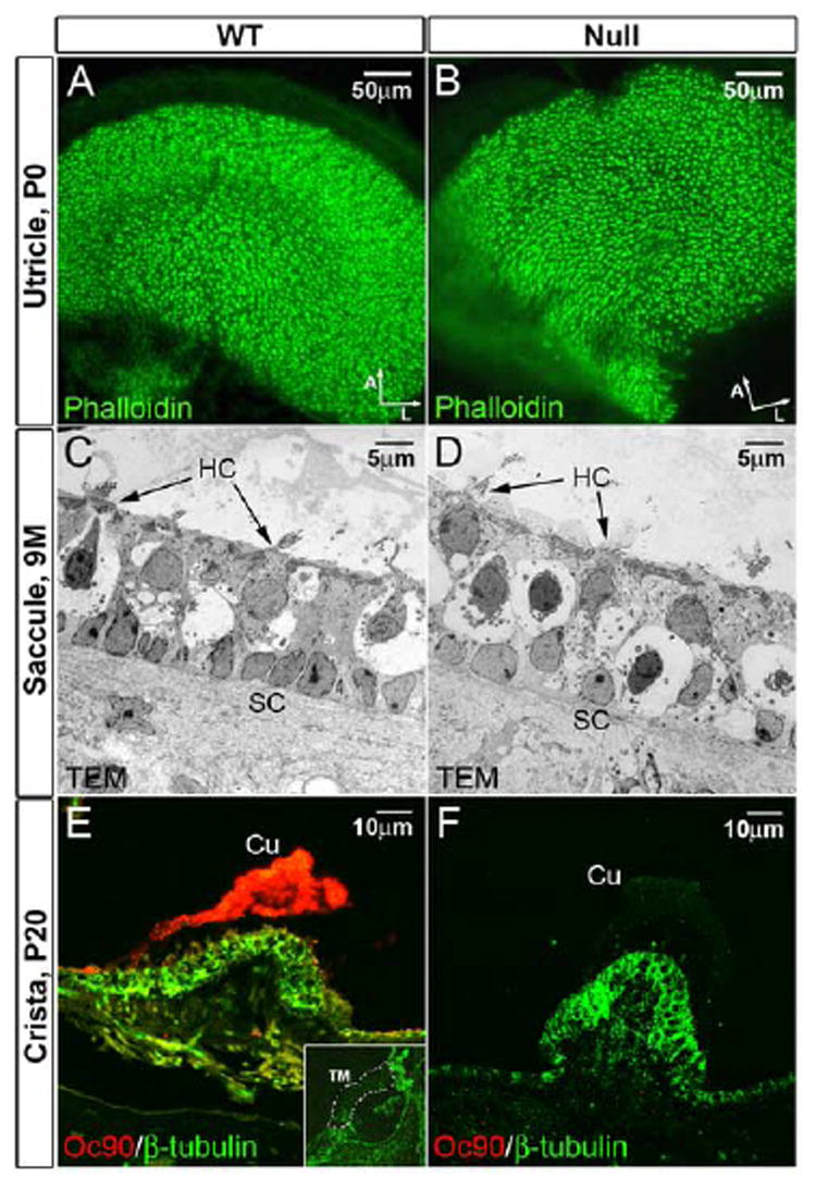
Figure 8.

(A, B) Normal density, organization and appearance of stereocilia in the Oc90 null vestibule as demonstrated by phalloidin-stained whole-mount utricle (P0). Arrows indicate orientation of whole-mount tissues (A, anterior; L, lateral). (C, D) Normal ultrastructure of the macular epithelial cells in Oc90 null mice (9 months old) as demonstrated by transmission electron microscopy. The amorphous gel-like structure above the macular sensory epithelium is also normal in the Oc90 null vestibule (only lightly visible in D due to the lighter background). (E, F) Oc90 is abundantly present in the wt cupula (P20 shown) but not in the null tissue. Shown in inset in (E) is the absence of Oc90 in the P20 wt tectorial membrane (delineated by dotted lines). The E16.5 wt tectorial membrane also has negative Oc90 signals. Cu, cupula; HC, hair cells; SC, supporting cells; TM, tectorial membrane.
