
Fig. 5.
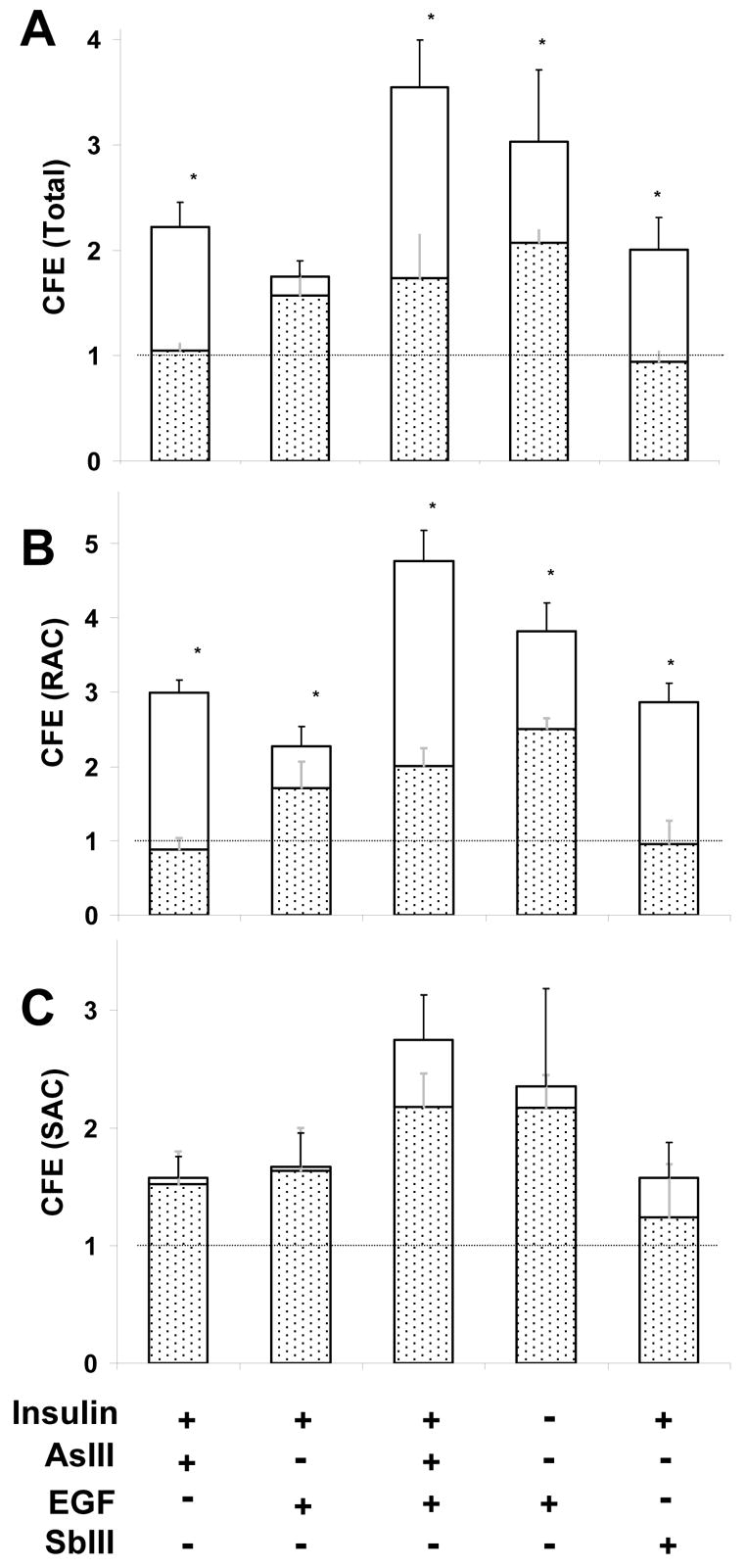
Effect of dominant negative β-catenin on proliferative potential of treated SIK. Cultures were treated as indicated for 3 days starting at 90% confluence before measuring CFE in (A) unfractionated, (B) rapidly adhering cell (RAC) and (C) slowly adhering cell (SAC) populations. The total bar height shows the response of cultures retrovirally infected with vector only (pBabe), and the stippled region shows the lower response of cultures infected with the T3 dominant negative β-catenin construct. Values are relative to untreated cultures in medium with insulin, set at 1. Significant differences (*) between pBabe and T3 infected SIK for indicated treatment are p<0.01 (Student’s t-test with Bonferroni correction) for three experiments in duplicate.