
Figure 1. apkcm/z mutants gastrulate but display progressive epithelial breakdown.
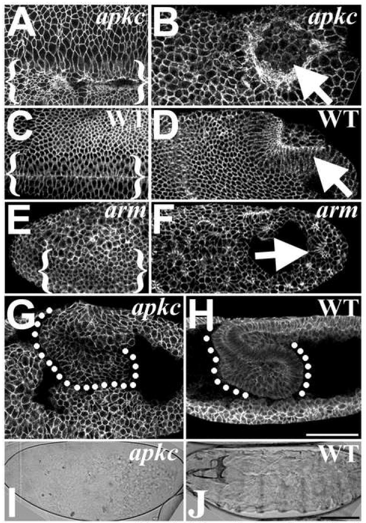
(A-H) Dlg labels cell cortexes. (A) apkcm/z mutant and (C) WT ventral furrows (bracketed). (B) apkcm/z mutant and (D) WT posterior midgut invaginations (arrows). (E) armm/z mutant ventral furrow (bracketed; furrow identified by Twist staining (data not shown)). (F) armm/z mutant posterior midgut invagination (arrow; embryo oriented by Miranda staining (data not shown)). The epithelium forms balls as it breaks down. (G) apkcm/z mutants undergo germband extension but then display widespread epithelial dissociation and cell rounding versus WT (H). (I) apkcm/z mutant cuticle—note small scraps of cuticle secreted from remaining epithelial cells. (J) WT. Bars, 50μm.