
Figure 5. MT asters are not released from centrosomes during apkc mutant cellularization.
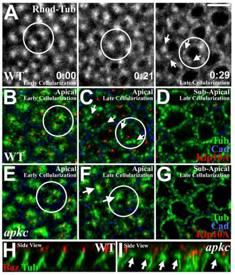
(A) Live imaging of WT embryos injected with rhodamine-labeled tubulin, focusing on apical MTs. Early cellularization (0:00, h:min): MTs form two apical asters per cell compartment. By late cellularization (0:29): apical asters break into MT bundles (arrows) running basally down the lateral membranes. (See Video 1) (B) Early WT cellularization. Apical MTs (green) are closely associated with two apical centrosomes (marked with Klp10a (red)) per cell compartment (plasma membrane: DE-Cad (blue)). (C) Late WT cellularization. Apical MTs (green) dissociate from centrosomes (red; arrows). (D) Imaging just basal to plane in (C) reveals lateral MT bundles in cross-section (green). (E) Early apkcm/z mutant cellularization. Apical MTs (green) associate with apical centrosomes (Klp10a (red)) as in WT (B). (F) Late apkcm/z mutant cellularization. MT asters (green) remain associated with centrosomes (red). (G) Imaging just basal to plane in (F) reveals lateral MT bundles in cross-section (green). (H) Side view, late WT cellularization. Note loss of apical MT asters. (I) Side view, late apkcm/z mutant cellularization. Note abnormal persistence of apical MT asters. Bar, 5μm.