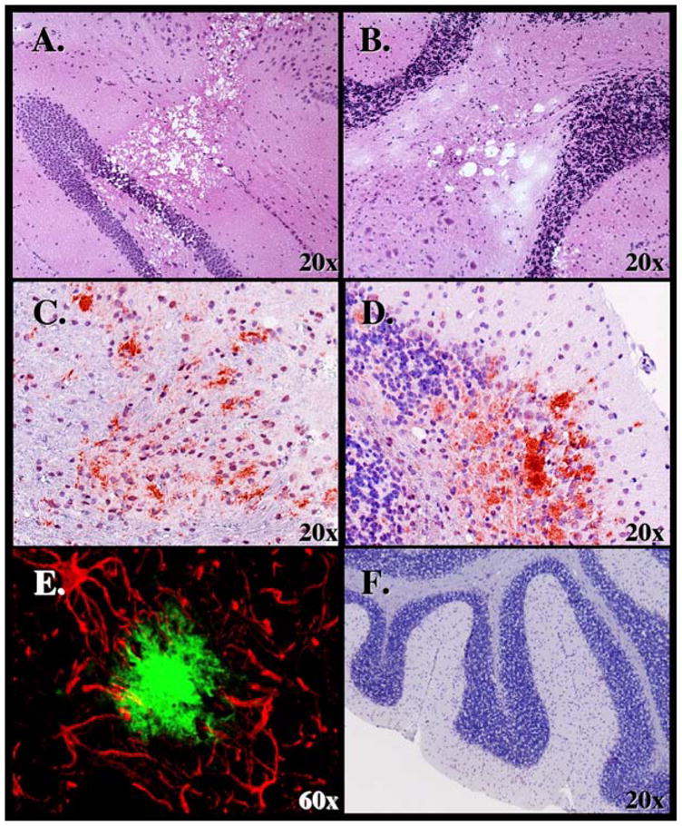
Figure 2.

Analysis of CNS tissues from deer PrP tg+/+ mice given CWD brain orally. Presence of spongiform lesions characteristic of TSE lesion in the hippocampus is shown in Panel A and cerebellum in Panel B. H & E staining used and mice sacrificed at 340 dpi. Panel C shows deposition of PrPres within the frontal cortex at 370 dpi and Panel D in the cerebellum at 320 dpi. D13 monoclonal antibody was used to detect PrPres for immunohistochemical analysis. Panel E displays double-immunochemical staining of an area in the cortex displaying astrocytic processes with antibody to GFAP (red) that surround a PrPres plaque (greenish/blue) stained with D13 antibody. Mouse sacrificed at 380 dpi. Panel F shows the absence of disease in cerebellum of deer PrP tg mouse inoculated with PBS diluent and sacrificed 400 dpi. Neither gliosis nor PrPres staining occurred in similar mice or in deer PrP tg mice inoculated with murine scrapie or non-tg mice inoculated with CWD.
