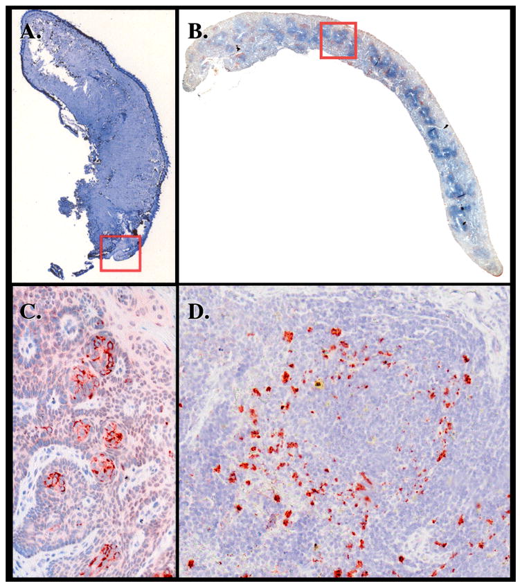
Figure 3.

Analysis of peripheral tissues from deer PrP tg+/+ mice given CWD brain orally. Panels A and C (insert from Panel A) are from the tongue of a mouse sacrificed 380 dpi. Serous and mucous glands in the posterior part of the dorsum of the tongue contain PrPres material detected with D13 antibody. Staining of same or adjacent section with monoclonal antibody to neuron-specific nuclear protein NeuN or with antibody to neurofilament H failed to show colocalization to PrPres (data not shown). This observation was confirmed in five additional deer PrP tg mice infected orally with deer PrP. Age-matched tongue specimens from deer PrP tg mice infected with murine scrapie or inoculated with PBS diluent or from C57Bl/6 mice inoculated with CWD brain were all negative (n = 5 or more/group). Panels B and D (insert of Panel B) are from the spleen. Panel D shows PrPres deposits in large cells, likely macrophage and/or dendritic cells, around splenic granule.
