
Figure 2.
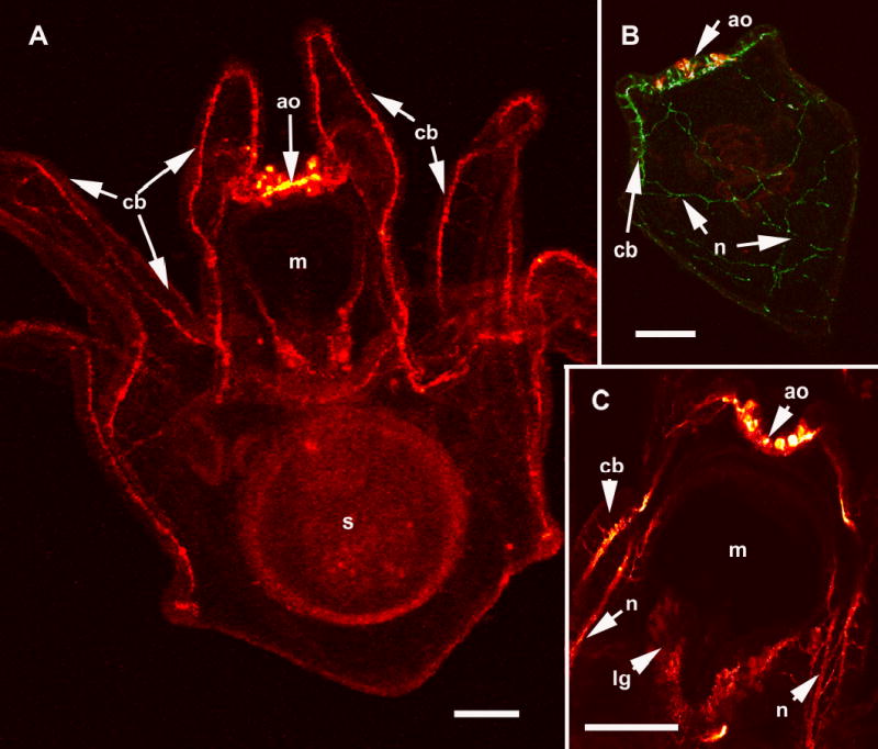
The larval nervous system of S. purpuratus revealed with immunolocalization of anti-synaptotagmin and anti-serotonin. A. Confocal image of an eight arm pluteus larva showing the tracts of axons that underlie the ciliated cells of the ciliary band (cb), anti-synaptotagmin. The apical organ (ao) is located on the oral hood at the foremost end of the larva and is thought to be a sensory organ. The larval mouth (m) is also innervated and nerves surround the esophagus, a tube connecting the mouth and the stomach (s). B. In the more familiar 4 arm pluteus larva the apical organ (ao) is double labelled with anti-serotonin (red) and the ciliary band neurons are labelled with anti-synaptotagmin (green). The neural cells bodies of the ciliary bands (cb) are arrayed at intervals along the oral side of the ciliated cells. The neurons in the ciliary bands project neurites to the posterior end of the larva (n). C. A confocal image (anti-synaptotagmin, red) showing details of the mouth (m) region. The lip ganglion (lg) contains serotonergic and non-serotonergic neurons. In addition to the ciliary band neurons and axon tracts (cb), there are bundles of neurites (n) of unknown function projecting from the apical organ, under the oral ectoderm. Bars = 25 μm