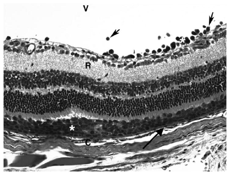
FIGURE 2.

Photomicrograph showing large Rev-2-T-6 lymphoma cells (*) localized in the subretinal space above the choroid (C) between the RPE (arrow) and retina (R). Some lymphoma cells (arrowhead) are also visible in the vitreous. Hematoxylin and eosin. Original magnification, ×400.
