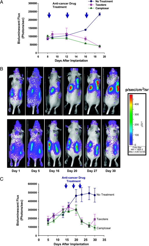
Figure 4.
Inhibition of the proliferation of mammary adenocarcinoma cells in hollow fibers in vivo by Taxotere and Camptosar. Nude mice bearing hollow fibers were either vehicle-treated or treated with Taxotere (20 mg/kg) or Camptosar (100 mg/kg) at indicated times (at 4-day intervals; arrowheads) after the implantation of hollow fibers filled with MAT B III-Luc-3H9 (A) or MCF7-Luc-10C11 (C) cells. Bioluminescence imaging was acquired longitudinally in these mice over the period shown. Data points show the means of six hollow fibers from three mice (each bearing two hollow fibers; error bars represent the standard error of the mean). (B) Longitudinal bioluminescence imaging of a representative mouse with either vehicle treatment (upper panel) or Taxotere treatment (lower panel) on days 15, 19 and 23, which harbors hollow fibers filled with MCF7-Luc-10C11 cells, was acquired on the days indicated after hollow fiber implantation.

