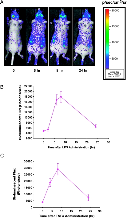
Figure 5.
Induction of NFκB reporter in hollow fibers in vivo by LPS and TNF-α. (A) Bioluminescence imaging of a representative mouse on pretreatment and posttreatment with LPS. (B and C) Nude mice harboring hollow fibers with MAT B III-NFκB-Luc cells were administered LPS (B; 2 mg/kg, i.p.) or TNF-α (C; 2 µg/mouse, i.p.). Bioluminescent images were acquired immediately before the administration of LPS or TNF-α, as well as at the indicated time after the administration of an antitumor agent. Data points show a mean bioluminescent flux (photons/sec) of six hollow fibers from three mice (each bearing two hollow fibers; error bars represent the standard error of the mean).

