
Figure 2.
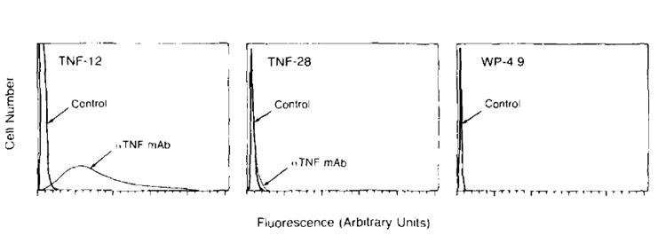
Flow microfluorimetric analysis of tumor cells stained with anti-human TNF antibody. A clone of the unmodified WP-4 parental culture. WP 4.9, as well as two TNF-producing clones, TNF-12 (high producer) and TNF-28 (low producer) were examined for the presence of membrane-associated human TNF. Fluorescence intensity was measured in arbitrary linear units. The 105 cells were analyzed in each frame. Negative controls stained with irrelevant antibody are represented by the sharp peaks to the far left. Cells staining positively for TNF are represented by the curves seen to the right of the controls. The high TNF producing clone was found to have significant amounts of membrane associated TNF (95% of the cells stained positively for TNF). In comparison, TNF-28, a low TNF secreter was found to express a very low amount of surface TNF. No evidence of membrane associated TNF was found on the nontransduced cell line WP 4.9.