
Fig. 5.
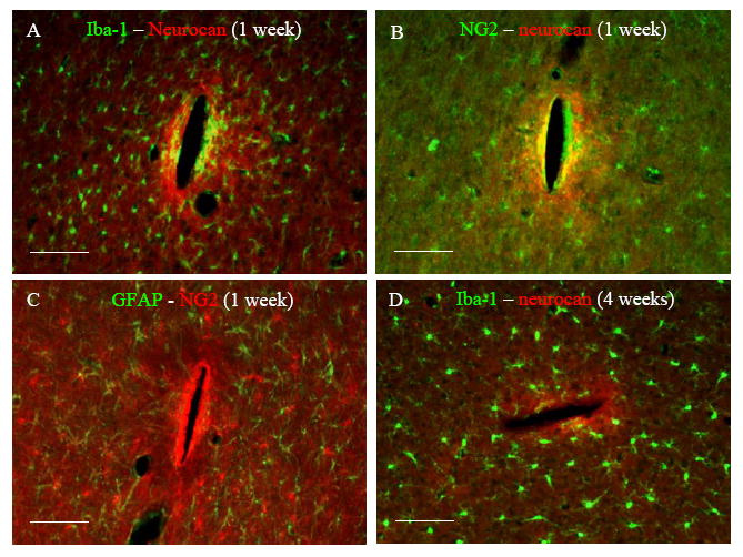
Representative fluorescent images of horizontal brain sections double stained with (A) Iba-1 – neurocan at 1 week, (B) NG2 – neurocan at 1 week, (C) GFAP – NG2 at 1 week, and (D) Iba-1 – neurocan 4 weeks post implantation. Scale bar = 100 μm.