
Figure 6.
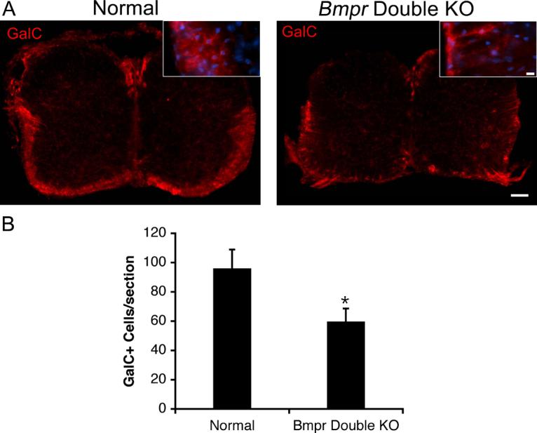
The number of cells expressing GalC is decreased in the Bmpr double knock out animals.
Spinal cords were removed from normal and Bmpr double knockout mice and processed for vibratome sectioning and immunolabeling with antibody to GalC. A) Shown are sections of whole spinal cord photographed at 10X and high magnification insets photographed at 40X. The scale bar for the 10X sections equals 100μm. The scale bar for the 40X insets equals 10μm. B) Cell counts performed at 20X magnification on three sections per animal from three animals taken from three separate litters. There is a 37% decrease in expression of GalC in the double knockout animals.