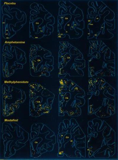
Figure 1.

Distribution of Fos-like immunoreactivity in the rostral brain of the cat. Camera lucida drawing of frontal sections showing Fos-ir neurons following oral administration of the indicated substances Note that (i) the placebo induces little labeling, (ii) amphetamine or methylphenidate treatment causes numerous Fos-positive neurons in the cerebral cortex and striatum, and (iii) modafinil treatment induces little labeling of the cortex and striatum, but a large number of aggregated positive neurons is seen in the anterior hypothalamic nucleus (AH). ACC, nucleus accumbens; CA, caudate nucleus; CL, claustrum; DBH, diagonal band of Broca; GP, globus pallidus; IC, internal capsule; LV, lateral ventricle; OC, optic chiasma; OLT, olfactory tubercle; OT, optic tract; Para, anterior paraventricular nucleus of the thalamus; PrF, prefrontal cortex; Pu, putamen; SI, substantia innominata; V3, third ventricle.
