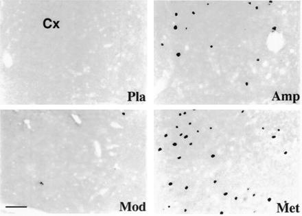
Figure 2.

Photomicrographs of frontal sections through the mediofrontal cortex (Cx) of the cat, showing Fos-like immunoreactivity following different administrations. Note that (i) the large number of stained cells seen with methylphenidate (Met) treatment and, to a lesser degree, with amphetamine (Amp) and (ii) few or no labeled neurons seen with modafinil (Mod) or placebo (Pla). (Bar = 50 μm.)
