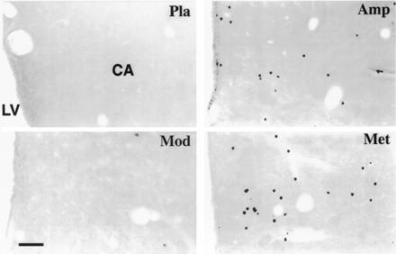
Figure 3.

Photomicrographs of frontal sections through the caudate nucleus (CA) of the cat, showing Fos-like immunoreactivity following different administrations. Note the large number of stained cells seen with amphetamine (Amp) or methylphenidate (Met) treatment and that few or no labeled neurons are seen using modafinil (Mod) or placebo (Pla). LV, lateral ventricle. (Bar = 80 μm.)
