Figure 1.

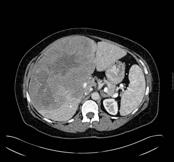
An axial CT image obtained during hepatic arterial phase demonstrates a heterogeneous enhancement of an approximately 22 × 14 cm hepatic mass involving the right hepatic lobe with a central region of hypoattenuation.

An axial CT image obtained during hepatic arterial phase demonstrates a heterogeneous enhancement of an approximately 22 × 14 cm hepatic mass involving the right hepatic lobe with a central region of hypoattenuation.