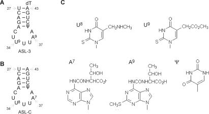
Figure 1.
ASL analogs of tRNALys and their modified bases. (A and B) Secondary structure presentations and indicated tRNA positions of ASL-3 and ASL-C, respectively. (C) Notations and structures of modified ASL bases. U9 (mcm5s2U), A9 (ms2t6A) appear in human tRNALys3, U8 (mnm5s2U) and A7 (t6A) in corresponding positions of E. coli tRNALys and Ψ appears in both.

