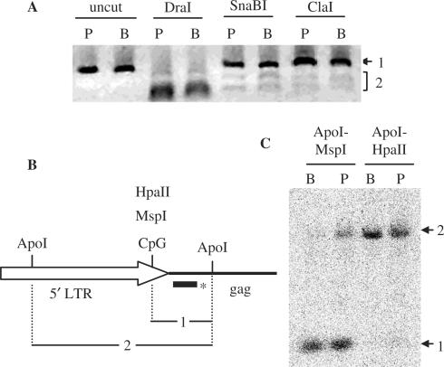
Figure 3.
Methylation of HERV-E and HERV-K sequences in placenta and PBL. (A) Genome-wide COBRA on HERV-E LTRs. Bisulfite treated DNA from placenta (P lanes) and PBL (B lanes) were used as template for PCR with primers amplifying HERV-E LTRs. PCR amplicons were treated with three restriction enzymes (SnaBI, ClaI and DraI). Bands 2 for SnaBI and ClaI digests correspond to sequences with methylated CpGs, whereas band 1 to a mix of sequences with unmethylated CpGs and sequences lacking the CpG site. The CpG free DraI restriction site is present in all sequences and was used as a control (see text). (B) and (C) methylation of HERV-K family determined by Southern blot. (B) Schematic representation of HERV-K 5′ LTR and gag region, with the restriction sites of the enzymes used. The bar with the asterisk corresponds to the probe overlapping the gag region. (C) Autoradiogram of the Southern blot. Lanes B are loaded with genomic DNA from PBL and lanes P with genomic DNA from placenta. Bands labeled 1 and 2 correspond to restriction fragments 1 and 2 shown in B.

