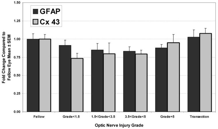
Figure 7.

Expression levels of two other proteins associated with differentiated astrocytes, GFAP and connexin43, were not altered in the ONH by either elevated IOP or transection. Although the pattern of expression in the various optic nerve injury groups may appear similar to that seen for TGFβ2 and aquaporin-4, levels in the optic nerve injury groups were not significantly different from fellow eye levels. Regression analysis also indicated no significant correlation.
