
Figure 1.
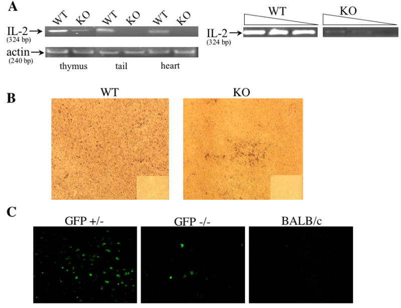
Detection of IL-2 DNA and IL-2 producing cells in IL-2 KO offspring of IL-2 heterozygous mothers. (A) Analysis of IL2 WT DNA in IL-2 KO mice. DNA was extracted from the thymus, heart, and tail of DO11.10/IL-2 WT or DO11.10/IL-2 KO mice and 300 ng were then used to detect the presence/absence of IL2 WT gene via PCR. β-actin was used as a loading control. In a separate experiment, DNA extracted from either DO11.10/IL-2 WT or DO11.10/IL-2 KO thymuses was serially diluted (undiluted, 1:10, 1:50) then assayed via PCR. (B) Thymic tissue sections from DO11.10/IL-2 KO offpring of DO11.10/IL-2 heterozygous mothers were processed for in situ hybridization, and IL-2 message was detected using a digoxigenin-labeled oligonucleotide probe cocktail for murine IL-2 mRNA (magnification 10x). The corresponding sense controls are shown in the right lower quadrant of each picture. (C) Thymic tissue sections from GFP−/− offspring of GFP+/− mothers expressing a transgenic IL-2 promoter/GFP reporter were assessed for the presence of GFP+ cells (magnification 10x). Sections from BALB/c mice were used as a negative control. Data shown in each figure are representative of several sections from 2 different animals.