
Figure 2.
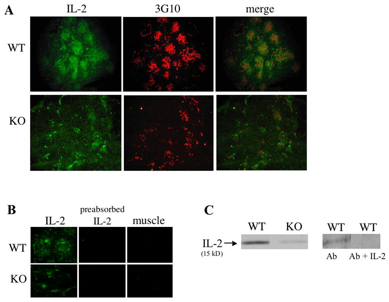
Detection of IL-2 protein in IL-2 WT and KO thymuses. (A) Cryosections of DO11.10/IL-2+/+ or DO11.10/IL-2−/− thymuses were stained for IL-2 (fluorescein isothiocyanate) and medullary epithelium (tetramethylrhodamine isothiocyanate) using clone 3G10 (magnification 10x). (B) Cryosections of DO11.10/IL-2+/+ or DO11.10/IL-2−/− thymuses or muscle were stained with a rat anti-mouse monoclonal antibody recognizing IL-2. To ensure specificity of the antibody, additional sections of thymus were stained with anti-IL-2 antibodies incubated with a 5M excess of IL-2 (preabsorbed IL-2). (C) Analysis of IL-2 protein by Western blot in DO11.10/IL-2 KO mice. Total protein was extracted from 1 thymic lobe of DO11.10/IL-2 WT or DO11.10/IL-2 KO mice, then separated by SDS-PAGE, transferred to nitrocellulose, and probed with anti-IL-2 antibodies. Pre-absorption of the primary anti-IL-2 antibody (Ab) with an excess of recombinant murine IL-2 (Ab + IL-2) showed no labeling.