
Figure 7.
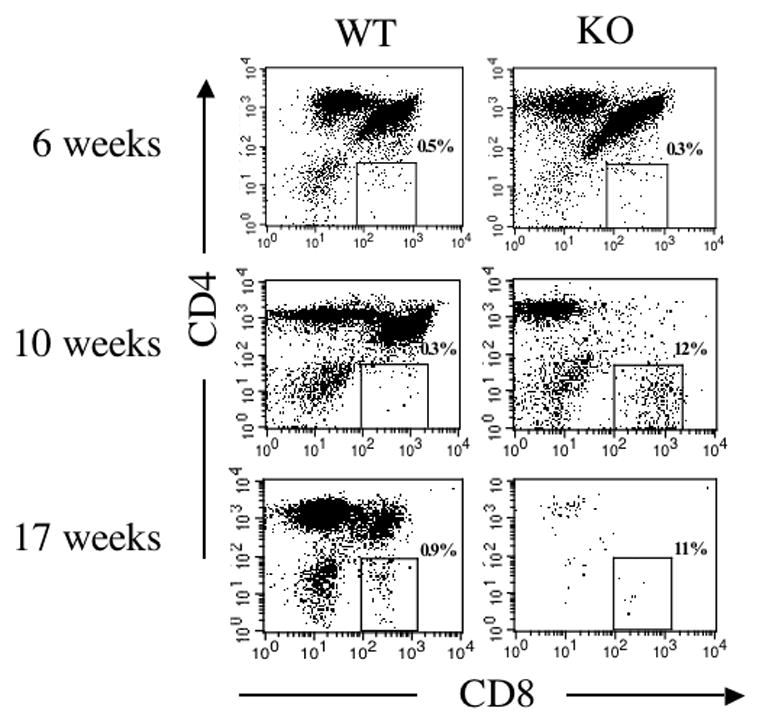
Generation of CD8+KJ1-26+ T cells in thymuses of RAG-1−/− mice reconstituted with either DO11.10/IL-2 WT or DO11.10/IL-2 KO bone marrow. Thymocytes, prepared from chimeras 6, 10, and 17 weeks post-transplantation, were assessed by flow cytometry for the presence of CD8+KJ1-26+ T cells. The dot plots shown represent the distribution of CD4+, CD8+, and CD4+CD8+ thymocytes within the KJ1-26+ thymocyte population. Data shown are representative of 3 or more experiments, with 3 animals per group.