
Figure 8.
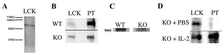
Activation of thymic lck in severe IL-2 deficiency. (A) Homogenates of whole thymuses were prepared using methods that maintained the phosphorylation state of thymic lck. Western blot analysis of the homogenates with an anti-lck antibody confirmed the presence of 56kD thymic lck. (B) Thymuses from RAG-1−/− mice, 12 weeks post-transplantation with either DO11.10/IL-2 WT or DO11.10/IL-2 KO bone marrow, were prepared as described above. Western blot analysis was then performed to assess the phosphorylation state of lck. Data are representative of 3 experiments, and extracts are from a single animal within one experiment. (C) Thymocyte lysates were prepared from 6 week-old DO11.10/IL-2 WT or KO thymuses. Lck was isolated from the lysates by anti-lck antibodies conjugated to agarose beads, and the phosphorylation state of lck was analyzed by Western blot. Densitometric analysis, performed with NIH Image software, revealed that the band generated from DO11.10/IL-2 WT mice was approximately 2.5 fold darker than that generated from DO11.10/IL-2 KO mice. Data are representative of 3 experiments, and extracts are from a single animal within one experiment. (D) DO11.10/IL-2 KO mice 10 – 15 weeks of age were treated with either PBS or 2 doses of IL-2 (1 μg/dose intraperitoneally, 24 h apart) and sacrificed 12 h later. Proteins were extracted and analyzed by Western blot as in A and B. Data are representative of 2 experiments, and extracts are from a single animal within one experiment.