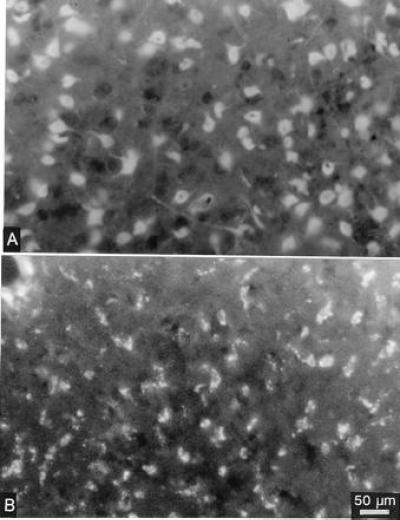
Figure 5.

Uptake of fluorescence labeled phosphorothioate oligonucleotides is shown 30 min (A) and 6 h (B) after a single injection (1 μl) into the striatum. The diffuse staining of cytoplasm, nuclei of nerve cell bodies, and their dendrites early after the injection changes to a granular appearance of the fluorescence at the 6-h time point. For Bregma level, see Fig. 4.
