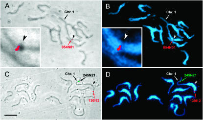
Figure 1.—
Tomato pachytene SC spreads viewed by phase contrast with superimposed FISH (A and C) and by UV illumination after DAPI staining and superimposed FISH (B and D). The dark spots observed on each SC by phase contrast are kinetochores, and the kinetochore of chromosome 1 is indicated in each image by an arrowhead. Pericentromeric heterochromatin stains more brightly with DAPI than distal euchromatic portions of the chromosomes. The locations of three BACs—054N01 (A and B, red signal), 0245N21 (C and D, green signal), and 130I12 (C and D, red signal) on chromosome 1—are indicated by arrows and text. The centromeric region of chromosome 1 has been enlarged (insets, A and B) to show that the FISH signal for 054N01 (marker SSR266) is located close to the centromere. Bar (A–D), 10 μm.

