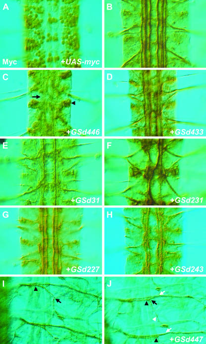
Figure 1.—
Selected examples of Fas2 phenotypes caused by misexpression of GS lines using scrt11-6-Gal4. (A) The nuclear targeted reporter UAS-nGFP, which also encodes five copies of the myc epitope, was used to demonstrate the expression driven by scrt11-6-Gal4 throughout the central and peripheral nervous systems of Drosophila embryos. Shown here is a dorsal view of three abdominal segments of an embryonic ventral nerve cord at stage 16, immunostained for myc. (B–J) Anti-Fas2 immunochemistry to reveal Fas2-positive axon bundles in the CNS (B–H) and motor neuron projections (I–J). (B) Three segments of a scrt11-6-Gal4/+ animal immunostained for Fas2, showing the characteristic Fas2-positive axon bundles traveling the length of the longitudinal connectives. (C) Misexpression of GSd446 causes many of the Fas2 bundles to become missing/disrupted or otherwise diffuse and less distinct (arrow). Fas2 staining was also mislocalized to the cell body (arrowhead). Additional phenotypes included inappropriate midline crossing, as in D–F and H, selective loss, or disruption of specific Fas2 axon bundles (E, G), hyperfasciculation (F), and misplaced axon bundles within the longitudinal connectives (F, H). (I) In the muscle field innervated by Fas2-positive motor neurons, SNa normally separates from the ISN at a point directly over the ventral muscle group (arrowhead), then bifurcates into two branches that innervate distinct muscle targets (black arrow). (J) Misexpression of GSd447 causes SNa to take an alternate trajectory that aligns with ISN for longer than usual (black arrowheads). Both the initial separation of SNa from ISN and the bifurcation of SNa (black arrow) appear intact, with a posterior-directed branch (white arrowhead) extending to muscle eight. However, after bifurcation the dorsal-directed branch of SNa often inappropriately rejoins ISN (white arrows), forming a fascicle that projects axons to more dorsal muscle targets.

