Figure 4.—
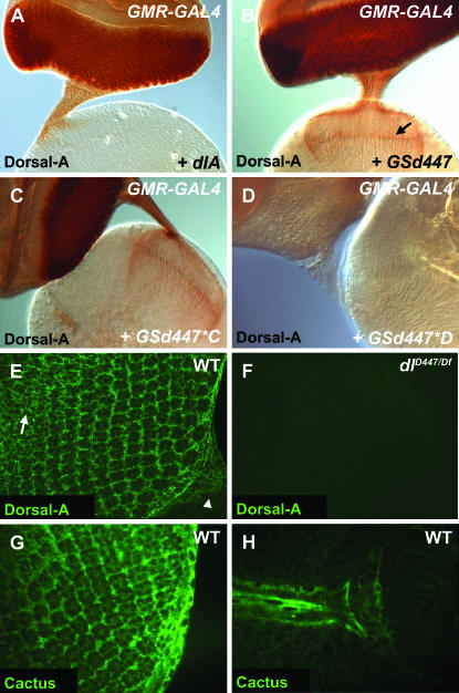
Dorsal and Cactus are expressed in the developing eye imaginal disc. α-Dorsal-A was used to stain the eye–brain complex (A–D). (A) Misexpression of UAS-dlA results in staining of the eye posterior to the morphogenetic furrow; however, little staining progresses past the optic stalk and into axon terminals (GMR-GAL4/UAS-dlA). (B) Misexpression of GSd447 resulted in intense staining for Dorsal-A in cell bodies and along entire axons to their terminals (GMR-GAL4/GSd447) (arrow). (C, D) The GSd447*C mutation remains immunoreactive for Dorsal (GMR-GAL4/GSd447*C) and retains localization to distal axons, while the GSd447*D mutation does not. (E) Confocal image of eye disc showing that Dorsal-A is expressed in the cell bodies of R cells in addition to cone cells and peripodial cells in wild-type (WT, genotype: w1118) eye discs. Anterior is upper left and posterior is lower right, with the beginning of the optic stalk marked by an arrowhead. The optical slice shown here largely cuts through a plane at the level of the R-cell nuclei. In more anterior positions, Dorsal-A is also expressed in undifferentiated cells prior to their recruitment into ommatidia (arrow). (F) In dl mutants (dlD447/Df(2L)TW119), expression is no longer observed, proving the specificity of the antibody staining pattern for Dorsal (F). (G, H) In WT (genotype: w1118) eye discs at third instar stages, Cactus was expressed in the cell bodies of photoreceptor neurons (G) in addition to cone cells, glial cells of the optic stalk (H), and peripodial cells.

