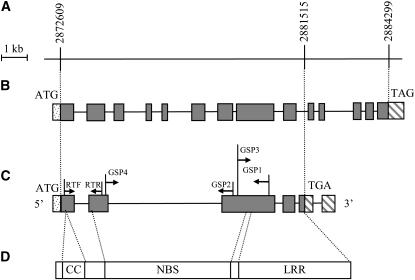
Figure 4.—
The structure of Pi36, as deduced from its genomic and cDNA sequences. (A) The target gene region. The numbers above the map show genomic positions in the Nipponbare genomic sequence. (B) Gene structure as deduced from the genomic DNA sequence. (C) Gene structure as deduced from the cDNA sequence. The shaded box indicates an exon, and the line an intron. The positions of 5′ and 3′ UTR (hatched boxes), the translation start codon (ATG), and the translation stop codon (TGA or TAG) are also shown. The annealing targets of the RACE and RT-PCR primers are indicated. (D) Structure of the Pi36-encoded protein, in which three tandem conserved domains are shown.

