
Fig. 2.
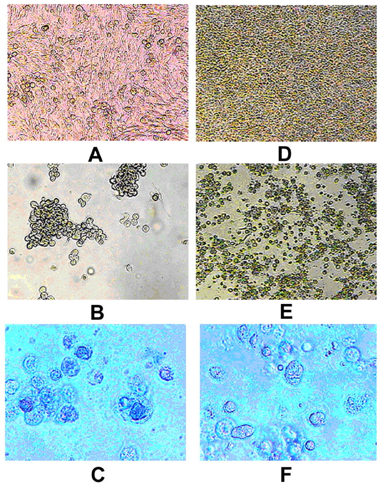
Morphology of mock infected and HSV-1 infected OCM1a and MUM2B melanoma cells. OCM1a and MUM2B cells were grown to approximately 70% confluency on 6-well tissue culture plates and were either exposed to 0.5 ml of sterile PBS (mock infection) or to HSV-1 diluted in PBS at a MOI=1. After incubation for 2 hours, the original inocula were removed and fresh tissue culture medium was added to each well and cultures were further incubated for times as indicated below when photographs were taken using an inverted light microscope. A. OCM1a culture 3 days after mock infection, B. OCM1a culture 3 days after HSV-1 infection, C. OCM1a culture 3 days after HSV-1 infection and following incubation of cultures with Trypan blue (0.2%) for 10 minutes, D. MUM2B culture 5 days after mock infection, E. MUM2B culture 5 days after HSV-1 infection, F. MUM2B culture 5 days after HSV-1 infection and following incubation of cultures with Trypan blue (0.2%) for 10 minutes.