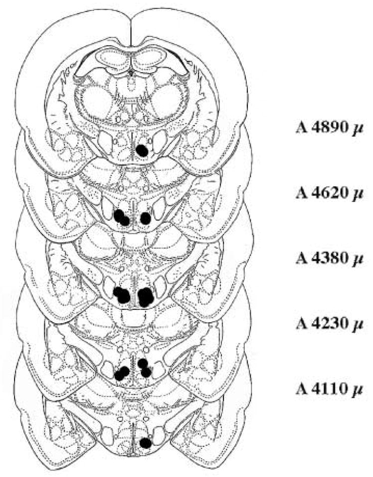
Figure 4. Location of cannulae from rats preinfused with 10−1 nmol BIM 90 min before 8-OH-DPAT or 8-OH-DPAT plus DOI.

The figure represents coronal sections taken from König and Klippel [16]at the level of the VMN from A 4110 μ to A 4890 μ. Dots on the left side of the sections indicate locations of bilateral cannulae from rat infused with 200 ng 8-OH-DPAT. Dots on the right represent locations of bilateral cannulae from rats infused with 200 ng 8-OH-DPAT and 2000 ng DOI.
