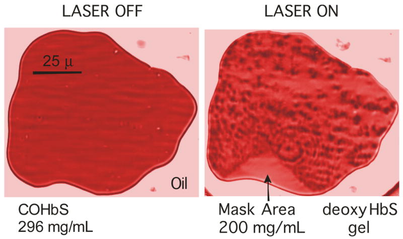
Figure 1.

A droplet of sickle hemoglobin in castor oil is shown before and after photolysis. The sample of chromatogrpahically purified Hb (in 0.15 M phosphate buffer at pH 7.35; 0.55 M sodium dithionite) has a thickness of 3.7 μm and is observed using a Leitz 32×LWD microscope objective; images are shown at 426 nm in the intensely absorbing Soret band. (Color has been introduced to help distinguish the regions). Laser photolysis by the 488 nm line of an Argon ion laser deoxygenates the COHb, which forms polymers. The rough texture is the result of the combination of turbidity, linear dichroism and non-uniformity of the polymer mass. On the perimeter of the droplet a triangular region has been masked from laser illumination, and remains HbCO, retaining the good optical characteristics of the unpolymerized Hb. Monomers diffuse from that region into the polymerized region as polymerization proceeds, until the final concentration is reached, as shown.
