
Figure 2.
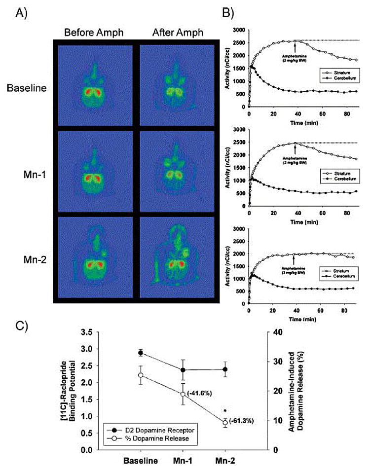
Fig. 2. Effect of chronic Mn exposure on [11C]-raclopride BP and AMPH-induced dopamine release. (A) Representative pseudocolor trans-axial images of [11C]-raclopride binding to D2-DAR in the striatum of one animal at baseline and at the Mn-1 and Mn-2 time points. Red areas represent high levels of binding and green areas represent low levels of [11C]-raclopride binding to D2-DAR. Note the progressive lack of a change in [11C]-raclopride levels in the striatum after AMPH administration from baseline to Mn-2. (B) [11C]-raclopride time–activity curves in the striatum and cerebellum before and after AMPH in the same animal as in panel A. Each graph corresponds to the adjacent images in panel A. At baseline (top graph in B), there is a dramatic decrease in [11C]-raclopride levels in the striatum after AMPH administration. Increasing Mn exposure reduces the effectiveness of AMPH-induced DA released to displace [11C]-raclopride from the striatum (see middle and lower graph in B). (C) Quantitative data on [11C]-raclopride BP and AMPH-induced in vivo DA release for all animals. For dopamine release, the numbers in parenthesis are the mean percent change from baseline. Each value is the mean ± SEM of 4 Mn-exposed animals. *p < 0.05. (Reprinted from: Nigrostriatal dopamine system dysfunction and subtle motor deficits in manganese-exposed non-human primates Experimental Neurology 202:381-390, 2006 with permission from Elsevier).