
Figure 5.
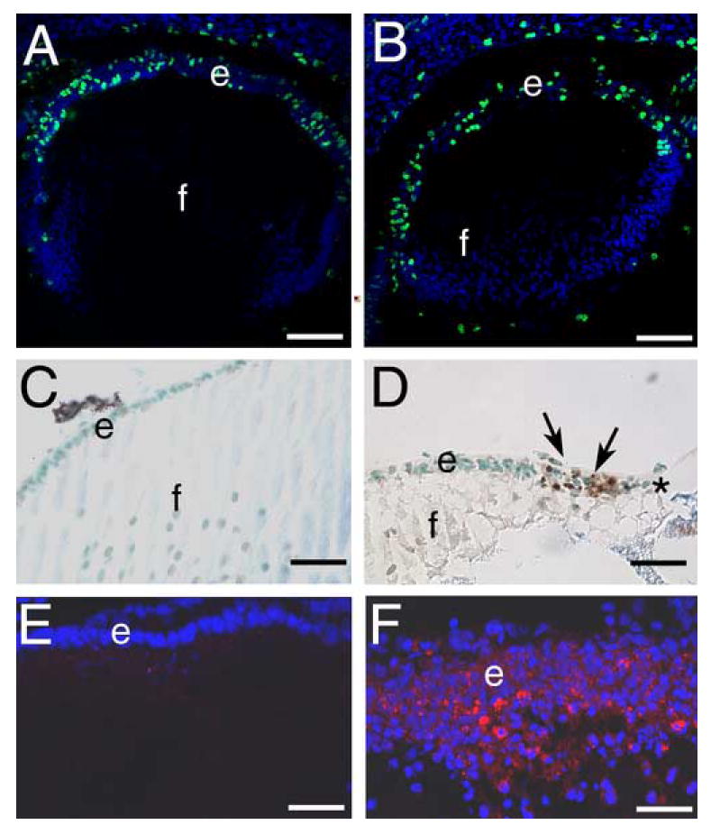
β1-integrin null lens epithelial cells are lost by birth due to elevated levels of apoptosis A, B) 16.5 dpc embryonic lenses stained for cells actively synthesizing DNA (green) using the 5-bromodeoxyuridine assay. Note that wildtype (A) and β1-integrin null (B) lens epithelia have qualitatively similar levels of cell proliferation. C, D) newborn lenses stained for apoptotic cells (brown) using TUNEL assay. Note that wildtype lenses (C) do not exhibit TUNEL positive lens epithelial cells while numerous TUNEL positive cells (arrows) are found in β1-integrin null lenses (D), especially in regions of lens epithelial cell loss (*). E, F) Newborn lens epithelial cells stained for cleaved caspase3 (Asp175). Note that wildtype lenses have little to no detectable cleaved caspase 3 (E) while numerous β1-integrin null lens epithelial cells are cleaved caspase 3 positive. Abbreviations: e- lens epithelium; f- lens fibers. Panels A,B: scale bar= 77 μm, C,D=30 μm, E,F=38 μm.